| Product Number |
ARP43628_P050 |
| Product Page |
www.avivasysbio.com/tap1-antibody-middle-region-arp43628-p050.html |
| Name |
TAP1 Antibody - middle region (ARP43628_P050) |
| Protein Size (# AA) |
808 amino acids |
| Molecular Weight |
87 kDa |
| NCBI Gene Id |
6890 |
| Host |
Rabbit |
| Clonality |
Polyclonal |
| Concentration |
0.5 mg/ml |
| Gene Full Name |
Transporter 1, ATP-binding cassette, sub-family B (MDR/TAP) |
| Alias Symbols |
APT1, PSF1, ABC17, ABCB2, PSF-1, RING4, TAP1N, D6S114E, TAP1*0102N |
| Peptide Sequence |
Synthetic peptide located within the following region: LVTFVLYQMQFTQAVEVLLSIYPRVQKAVGSSEKIFEYLDRTPRCPPSGL |
| Product Format |
Liquid. Purified antibody supplied in 1x PBS buffer with 0.09% (w/v) sodium azide and 2% sucrose. |
| Reference |
Soundravally,R. (2008) Scand. J. Immunol. 67 (6), 618-625 |
| Description of Target |
The membrane-associated protein encoded by this gene is a member of the superfamily of ATP-binding cassette (ABC) transporters. ABC proteins transport various molecules across extra- and intra-cellular membranes. ABC genes are divided into seven distinct subfamilies (ABC1, MDR/TAP, MRP, ALD, OABP, GCN20, White). TAP1 is a member of the MDR/TAP subfamily. Members of the MDR/TAP subfamily are involved in multidrug resistance. TAP1 is involved in the pumping of degraded cytosolic peptides across the endoplasmic reticulum into the membrane-bound compartment where class I molecules assemble.The membrane-associated protein encoded by this gene is a member of the superfamily of ATP-binding cassette (ABC) transporters. ABC proteins transport various molecules across extra- and intra-cellular membranes. ABC genes are divided into seven distinct subfamilies (ABC1, MDR/TAP, MRP, ALD, OABP, GCN20, White). This protein is a member of the MDR/TAP subfamily. Members of the MDR/TAP subfamily are involved in multidrug resistance. The protein encoded by this gene is involved in the pumping of degraded cytosolic peptides across the endoplasmic reticulum into the membrane-bound compartment where class I molecules assemble. Mutations in this gene may be associated with ankylosing spondylitis, insulin-dependent diabetes mellitus, and celiac disease. Publication Note: This RefSeq record includes a subset of the publications that are available for this gene. Please see the Entrez Gene record to access additional publications. |
| Protein Interactions |
UBC; MUL1; TDP2; SFXN1; GADD45GIP1; MRPL40; MCAT; SRSF3; SDHA; ABCD3; CYC1; PSMB8; TAPBP; TAP2; HLA-A; PDIA3; CALR; B2M; PDIA2; KRTAP4-12; MDFI; PSMB5; HLA-F; HLA-G; ESR1; COPG1; COPB1; |
| Reconstitution and Storage |
For short term use, store at 2-8C up to 1 week. For long term storage, store at -20C in small aliquots to prevent freeze-thaw cycles. |
| Enhanced Validation |
| Relative Expression (Western Blot) |
 |
|
| Datasheets/Manuals |
Printable datasheet for anti-TAP1 (ARP43628_P050) antibody |
| Blocking Peptide |
For anti-TAP1 (ARP43628_P050) antibody is Catalog # AAP43628 (Previous Catalog # AAPP11617) |
| Immunogen |
The immunogen is a synthetic peptide directed towards the middle region of human TAP1 |
| Uniprot ID |
Q03518 |
| Protein Name |
Antigen peptide transporter 1 |
| Sample Type Confirmation |
TAP1 is supported by BioGPS gene expression data to be expressed in HeLa |
| Protein Accession # |
NP_000584 |
| Purification |
Affinity Purified |
| Nucleotide Accession # |
NM_000593 |
| Tested Species Reactivity |
Human |
| Gene Symbol |
TAP1 |
| Predicted Species Reactivity |
Human, Mouse, Rat, Cow, Guinea Pig, Horse, Rabbit, Zebrafish |
| Application |
IHC, WB |
| Predicted Homology Based on Immunogen Sequence |
Cow: 92%; Guinea Pig: 86%; Horse: 93%; Human: 100%; Mouse: 100%; Rabbit: 92%; Rat: 100%; Zebrafish: 83% |
| Image 1 | Human Ovary, HepG2
 | Host: Rabbit
Target: TAP1
Positive control (+): Human Ovary (OV)
Negative control (-): HepG2 (HG)
Antibody concentration: 4ug/ml |
|
| Image 2 | Human Lung Tissue
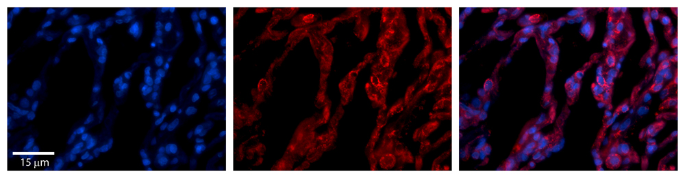 | Rabbit Anti-TAP1 Antibody
Catalog Number: ARP43628_P050
Formalin Fixed Paraffin Embedded Tissue: Human Lung Tissue
Observed Staining: Cytoplasmic in alveolar type I & II cells
Primary Antibody Concentration: 1:100
Secondary Antibody: Donkey anti-Rabbit-Cy3
Secondary Antibody Concentration: 1:200
Magnification: 20X
Exposure Time: 0.5 - 2.0 sec
|
|
| Image 3 | Western Blot
 | 25 ug of the indicated Human whole cell extracts was loaded onto a 12% SDS-PAGE gel. 4 ug/mL of the antibody was used in this experiment. Recommended dilution for this antibody is 1-3 ug/mL. |
|