- Toll Free: 888-880-0001
- Phone: 858-552-6979
- Email: info@avivasysbio.com
- $55: Antibody & Protein in US
- $55 + $25/Kit in US
- Contact us for international orders.

| Datasheets/Manuals | Printable datasheet for anti-NME1 (AVARP00009_P050) antibody |
|---|
| Tested Species Reactivity | Human |
|---|---|
| Predicted Species Reactivity | Human, Mouse, Rat, Cow, Guinea Pig, Horse, Rabbit |
| Product Format | Liquid. Purified antibody supplied in 1x PBS buffer with 0.09% (w/v) sodium azide and 2% sucrose. |
| Clonality | Polyclonal |
| Host | Rabbit |
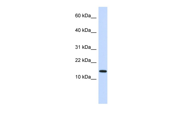
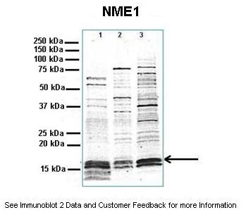
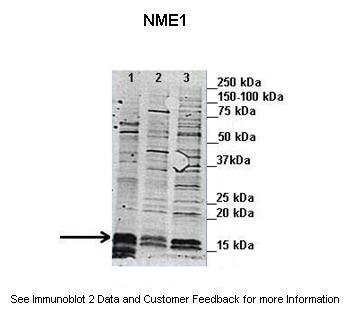
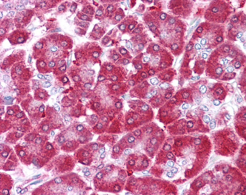
| Application | DB, IHC, WB |
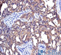
| Additional Information | IHC Information: Immunohistochemical analysis of paraffin-embedded human lung adenocarcinoma tissue using anti-Nm23-H1 antibody (AVARP00009_P050). IHC Information: Pancreas IHC Information: Small intestine |
| Reconstitution and Storage | For short term use, store at 2-8C up to 1 week. For long term storage, store at -20C in small aliquots to prevent freeze-thaw cycles. |
| Immunogen | The immunogen is a synthetic peptide directed towards the N terminal region of human NME1 |
| Purification | Affinity Purified |
| Predicted Homology Based on Immunogen Sequence | Cow: 100%; Guinea Pig: 92%; Horse: 100%; Human: 100%; Mouse: 100%; Rabbit: 100%; Rat: 100% |
| Peptide Sequence | Synthetic peptide located within the following region: ANCERTFIAIKPDGVQRGLVGEIIKRFEQKGFRLVGLKFMQASEDLLKEH |
| Concentration | 0.5 mg/ml |
| Blocking Peptide | For anti-NME1 (AVARP00009_P050) antibody is Catalog # AAP30457 (Previous Catalog # AAPP01041) |
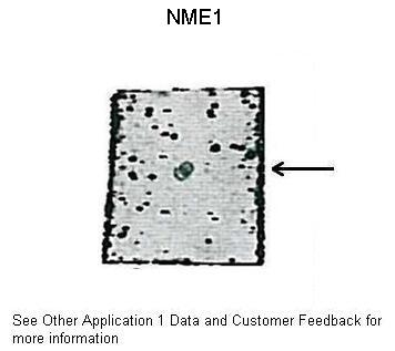
| Other Applications Image 1 Data | DB Suggested Anti-Nme1 antibody Titration: 0.5 ug/ml Positive Control: Recomninant human NME2 protein |
| Reference | Liu,S.J., (2008) Br. J. Cancer 98 (2), 363-369 |
| Gene Symbol | NME1 |
|---|---|
| Gene Full Name | Non-metastatic cells 1, protein (NM23A) expressed in |
| Alias Symbols | NB, AWD, NBS, GAAD, NDKA, NM23, NDPKA, NDPK-A, NM23-H1 |
| NCBI Gene Id | 4830 |
| Protein Name | Nucleoside diphosphate kinase B |
| Description of Target | This gene (NME1) was identified because of its reduced mRNA transcript levels in highly metastatic cells. Nucleoside diphosphate kinase (NDK) exists as a hexamer composed of 'A' (encoded by this gene) and 'B' (encoded by NME2) isoforms. Mutations in this gene have been identified in aggressive neuroblastomas. Two transcript variants encoding different isoforms have been found for this gene. Co-transcription of this gene and the neighboring downstream gene (NME2) generates naturally-occurring transcripts (NME1-NME2), which encodes a fusion protein comprised of sequence sharing identity with each individual gene product.This gene (NME1) was identified because of its reduced mRNA transcript levels in highly metastatic cells. Nucleoside diphosphate kinase (NDK) exists as a hexamer composed of 'A' (encoded by this gene) and 'B' (encoded by NME2) isoforms. Mutations in this gene have been identified in aggressive neuroblastomas. Two transcript variants encoding different isoforms have been found for this gene. Co-transcription of this gene and the neighboring downstream gene (NME2) generates naturally-occurring transcripts (NME1-NME2), which encodes a fusion protein comprised of sequence sharing identity with each individual gene product. |
| Uniprot ID | P22392 |
| Protein Accession # | NP_000260 |
| Nucleotide Accession # | NM_000269 |
| Protein Size (# AA) | 152 |
| Molecular Weight | 17kDa |
| Protein Interactions | SREK1IP1; PID1; TNPO2; POLR1C; NME4; NME3; NME1; TUBG1; UBC; MDM2; ASB18; BMI1; CHRAC1; NME1-NME2; WDR1; THOP1; SORD; RBBP7; OXCT1; IDH1; FDPS; MAPK14; FBXO6; WDYHV1; NIF3L1; SOX30; FXR2; SMARCD1; NME2; CCND3; UROD; SOD1; MIF; ERG; EBNA-LP; HDAC5; STRAP; |
- Protocol:
- Reconstitution & Storage Instructions
- Western Blotting/Immunoblotting (WB/IB) Protocol
- Immunohistochemistry (IHC) Protocol
- Immunocytochemistry (ICC) Protocol
- Enzyme-Linked ImmunoSorbent Assay (ELISA) Protocol
- Blocking Peptide Competition Protocol (BPCP)
- Immunoprecipitation (IP) Protocol
- Antibody Array (AA) Protocol
- Tips Information:
-
See our General FAQ page.
-
What is the species homology for "NME1 Antibody - N-terminal region (AVARP00009_P050)"?
The tested species reactivity for this item is "Human". This antibody is predicted to have homology to "Human, Mouse, Rat, Cow, Guinea Pig, Horse, Rabbit".
-
How long will it take to receive "NME1 Antibody - N-terminal region (AVARP00009_P050)"?
This item is available "Domestic: within 1-2 days delivery | International: 1-2 days".
-
What buffer format is "NME1 Antibody - N-terminal region (AVARP00009_P050)" provided in?
This item is provided in "Liquid. Purified antibody supplied in 1x PBS buffer with 0.09% (w/v) sodium azide and 2% sucrose.".
Additional format options may be available. For more information please contact info@avivasysbio.com. -
What are other names for "NME1 Antibody - N-terminal region (AVARP00009_P050)"?
This target may also be called "NB, AWD, NBS, GAAD, NDKA, NM23, NDPKA, NDPK-A, NM23-H1" in publications.
-
What is the shipping cost for "NME1 Antibody - N-terminal region (AVARP00009_P050)"?
The shipping cost for this item is $40 within the US. Please contact us for specific shipping prices for international orders.
-
What is the guarantee for "NME1 Antibody - N-terminal region (AVARP00009_P050)"?
All Aviva products have been through rigorous validations and carry 100% satisfaction guarantee.
-
Can I get bulk pricing for "NME1 Antibody - N-terminal region (AVARP00009_P050)"?
You can get bulk pricing for this item by going here.
-
What is the molecular weight of the protein?
The molecular weight reported by Uniprot for this item is "17kDa".
Please note observed molecular weights in western blot applications may differ depending on a variety of protein characteristics. -
What protocols are available for "NME1 Antibody - N-terminal region (AVARP00009_P050)"?
We may have detailed protocol data avaialble for this item. To learn more, please view the "Protocols & Data" tab on the product page.
-
What are positive controls for "NME1"?
We have listed RNA Seq and gene expression data in the "Target Info" tab. You may be able to find adequate positive controls there.
-
What are negative controls for "NME1"?
We have listed RNA Seq and gene expression data in the "Target Info" tab. You may be able to find adequate positive controls there.
-
What other proteins interact with "NME1"?
This protein has been reported to interact with "Protein Interactions". Please view the "Related Categories" tab on the product page for more information.
-
What biological processes are associated with "NME1"?
This protein has been associated with "Biological Processes". Please view the "Related Categories" tab on the product page for more information.
-
What cellular components are associated with "NME1"?
This protein has been associated with "Cellular Components". Please view the "Related Categories" tab on the product page for more information.
-
What protein functions are associated with "NME1"?
This protein has been associated with "Protein Functions". Please view the "Related Categories" tab on the product page for more information.


